Prenatal Diagnosis
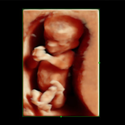
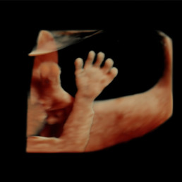
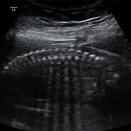
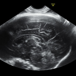
The best way to know your baby's health is through special studies called genetic or morphologic (or second level) ultrasounds. There is one for each trimester and each one detects different information specific to that trimester. This kind of special ultrasounds are performed by certified and trained physicians called fetal maternal specialists.
The time to do these ultrasounds and the objectives of each of them are:
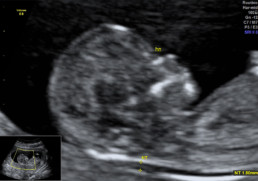
This ultrasound allows to determine the risk of trisomy 21 (Down syndrome), trisomy 18 (Edwards syndrome) or trisomy 13 (Patau syndrome) with the measurement of certain ultrasound markers like nuchal translucency, nasal bone, tricuspid regurgitation and ductus venosus; as well as an early inspection of the fetal anatomy. If higher risk of preeclampsia or intrauterine growth restriction is detected, medication can be given to reduce and treat the disorder.
- Confirm intrauterine gestation.
- Determine the number of fetuses and placentas.
- Adequately date the gestation.
- Determination of markers of certain chromosomal defects.
- Early anatomical inspection.
- Establish the risk of preeclampsia, premature birth, and intrauterine growth restriction.
This study is crucial because it permits detection of fetal malformations, for example, cleft lip palate, approximately 80% of congenital heart defects, open spina bifida, certain brain abnormalities and many others. If timely diagnosis is done, some can be treated in utero and others, at birth; thefore, allowing a better prognosis and quality of life for the baby.
- Evaluate fetal anatomy.
- Establish preeclampsia risk.
- Determine likelihood of premature birth.
- Evaluation of placenta, fetal cord insertion and amniotic fluid.
- Estimate fetal growth.
This sonogram is important as it provides an additional checkup of the fetal structure. Some diseases such as microcephaly and intrauterine growth restriction (IUGR) are prone to develop late in pregnancy. Detection and follow up of IUGR can greatly reduce the rate of morbidity and mortality of this fetal/placental disease. This can be found in about 10% of the pregnancies.
- Estimate fetal growth.
- Rescan of fetal anatomy to detect diseases that progress or appear late in pregnancy.
- Evaluate fetal movements.
- Check for placenta and amniotic fluid.
- Establish preeclampsia risk.
In certain cases, additional ultrasounds may be needed. For example: multiple pregnancy, diseases of pregnancy like diabetes or hypertension, etc.
4. Fetal echocardiography
This study offers a more detailed and in depth checkup of the fetal heart. Some indicators that may require this process include but are not limited to mothers with a congenital heart disease, suspected congenital heart disease in the baby, fetus with a reversed a-wave ductus venosus in first trimester, among many others.
6. Biophysical profile
This is considered a fetal well-being test that evaluates different parameters in the baby, like breathing movements, gross body movements, muscular tone, amniotic fluid index and non-stress test. The first 4 parameters are seen and measured in an ultrasound and the last test is done by cardiotocography to monitor the baby’s heart rate and contractions.


